


 Foundational characteristics of cancer include proliferation, angiogenesis, migration, evasion of apoptosis, and cellular immortality. Find key markers for these cellular processes and antibodies to detect them.
 The SUMOplot™ Analysis Program predicts and scores sumoylation sites in your protein. SUMOylation is a post-translational modification involved in various cellular processes, such as nuclear-cytosolic transport, transcriptional regulation, apoptosis, protein stability, response to stress, and progression through the cell cycle.
 The Autophagy Receptor Motif Plotter predicts and scores autophagy receptor binding sites in your protein. Identifying proteins connected to this pathway is critical to understanding the role of autophagy in physiological as well as pathological processes such as development, differentiation, neurodegenerative diseases, stress, infection, and cancer.
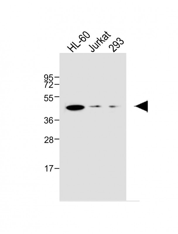
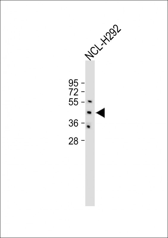
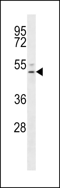
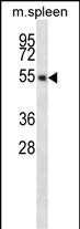
EDG6 Antibody (N-term)
Affinity Purified Rabbit Polyclonal Antibody (Pab)
- SPECIFICATION
- CITATIONS: 2
- PROTOCOLS
- BACKGROUND

Application
| WB, E |
|---|---|
| Primary Accession | O95977 |
| Other Accession | NP_003766 |
| Reactivity | Human, Mouse |
| Host | Rabbit |
| Clonality | Polyclonal |
| Isotype | Rabbit IgG |
| Calculated MW | 41623 Da |
| Antigen Region | 8-37 aa |
| Gene ID | 8698 |
|---|---|
| Other Names | Sphingosine 1-phosphate receptor 4, S1P receptor 4, S1P4, Endothelial differentiation G-protein coupled receptor 6, Sphingosine 1-phosphate receptor Edg-6, S1P receptor Edg-6, S1PR4, EDG6 |
| Target/Specificity | This EDG6 antibody is generated from rabbits immunized with a KLH conjugated synthetic peptide between 8-37 amino acids from the N-terminal region of human EDG6. |
| Dilution | WB~~1:1000 E~~Use at an assay dependent concentration. |
| Format | Purified polyclonal antibody supplied in PBS with 0.09% (W/V) sodium azide. This antibody is purified through a protein A column, followed by peptide affinity purification. |
| Storage | Maintain refrigerated at 2-8°C for up to 2 weeks. For long term storage store at -20°C in small aliquots to prevent freeze-thaw cycles. |
| Precautions | EDG6 Antibody (N-term) is for research use only and not for use in diagnostic or therapeutic procedures. |
| Name | S1PR4 |
|---|---|
| Synonyms | EDG6 |
| Function | G protein-coupled receptor highly expressed in immune cells, where it regulates immune response and cytokine production. Functions as a receptor for the lysosphingolipid sphingosine-1-phosphate (S1P). Upon S1P binding, promotes regulatory T-cell differentiation and enhances fatty acid oxidation, through activation of the NRF2/PPARA signaling pathway (By similarity). Modulates also M1 macrophage activation through interaction with FPR2 and the JNK signaling, contributing to the inflammatory response (By similarity). In addition, facilitates early neutrophil mobilization and vascular activation during inflammation, promoting lymphocyte recruitment to draining lymph nodes and supporting the development of germinal centers for an effective adaptive immune response (By similarity). |
| Cellular Location | Cell membrane; Multi-pass membrane protein. |
| Tissue Location | Specifically expressed in fetal and adult lymphoid and hematopoietic tissue as well as in lung. Considerable level of expression in adult and fetal spleen as well as adult peripheral leukocytes and lung. Lower expression in adult thymus, lymph node, bone marrow, and appendix as well as in fetal liver, thymus, and lung |

Provided below are standard protocols that you may find useful for product applications.
Background
EDG6 is a member of the G protein-coupled receptors, as well as the EDG family of proteins. It participates in endothelial differentiation, and may regulate lymphocyte cell signaling. It is a member of the lysophospholipid/lysosphingolipid receptor family.
References
Contos, J.J., et al., FEBS Lett. 531(1):99-102 (2002).
Yamazaki, Y., et al., Biochem. Biophys. Res. Commun. 268(2):583-589 (2000).
Graler, M.H., et al., Genomics 53(2):164-169 (1998).
If you have used an Abcepta product and would like to share how it has performed, please click on the "Submit Review" button and provide the requested information. Our staff will examine and post your review and contact you if needed.
If you have any additional inquiries please email technical services at tech@abcepta.com.
Ordering Information
Other Products
Shipping Information
